Summary
Discover_How_a_Specific_Type_of_Mammogram_Benefits_Women_with_Dense_Breasts explores the advantages of advanced mammographic techniques, particularly digital breast tomosynthesis (DBT) and contrast-enhanced mammography (CEM), in improving breast cancer detection among women with dense breast tissue. Dense breasts, which contain higher proportions of fibroglandular tissue relative to fat, appear opaque on traditional two-dimensional (2D) mammograms, often masking tumors that similarly appear white, thereby reducing the sensitivity and specificity of standard screening methods. Given that nearly half of women aged 40 and older have dense breasts, this presents a significant clinical challenge and increases the risk of missed or delayed breast cancer diagnoses.
Digital breast tomosynthesis, commonly known as 3D mammography, generates multiple cross-sectional images of the breast, reducing tissue overlap and enhancing lesion visibility. This technology has been shown to increase cancer detection rates and decrease false-positive recalls in women with dense breasts compared to conventional mammography. Contrast-enhanced mammography, which involves the intravenous administration of an iodine-based contrast agent, highlights tumor neovascularity and vascular permeability, further improving detection in dense tissue where traditional imaging often fails. Together, these modalities provide more accurate tumor characterization and staging, aiding clinical decision-making and potentially improving patient outcomes.
Despite these advances, challenges remain regarding accessibility, cost, and the optimal integration of these technologies into routine screening protocols. While DBT is increasingly recommended and supported by updated guidelines and legislation requiring breast density notification, the use of CEM is still emerging, with ongoing clinical trials assessing its role alongside DBT and other imaging methods. Additionally, the use of contrast agents in CEM introduces considerations about patient safety and procedure complexity.
Patient advocacy and legislative efforts have been pivotal in raising awareness of dense breast issues and accelerating the adoption of improved screening technologies like DBT. Future directions focus on combining advanced imaging with personalized risk assessment and exploring new screening paradigms to enhance early detection and reduce breast cancer mortality among women with dense breasts.
Background
Breast density refers to the proportion of fibroglandular tissue relative to fatty tissue within the breast, as seen on a mammogram. Dense breast tissue contains higher amounts of glandular and fibrous connective tissue and lower amounts of fat, making it appear opaque on imaging. Nearly half of women aged 40 and older who undergo mammography are found to have dense breasts. This density presents a challenge in breast cancer detection because tumors, which also appear opaque, can be obscured by the surrounding dense tissue, reducing the sensitivity and specificity of traditional mammography.
Traditional mammograms produce two-dimensional (2D) images of the breast, usually from two angles, but these can struggle to differentiate abnormalities within dense tissue. Dense breasts not only make tumors harder to detect but are also associated with an increased risk of breast cancer, although this does not necessarily translate to higher mortality after diagnosis once other factors are adjusted for. Breast cancer incidence rises with age and peaks between 70 and 74 years, with mortality also increasing with age; however, clinical trials have not definitively established the benefits of screening women aged 75 or older.
To overcome the limitations posed by dense breast tissue, imaging techniques such as 3D mammography (digital breast tomosynthesis) and contrast-enhanced mammography (CEM) have been developed. 3D mammography generates multiple cross-sectional images, improving lesion visibility and aiding in better sizing and staging of masses, which can reduce unnecessary recall rates in women with dense breasts. CEM uses an iodine-based intravenous contrast agent that highlights areas of increased vascularity, such as tumors with leaky vessels, allowing these regions to appear brighter on subtracted images and improving detection in dense tissue where traditional mammograms may miss abnormalities. However, benign background enhancement can sometimes limit sensitivity with CEM, similar to breast MRI. These advanced imaging modalities provide improved visualization of dense breast tissue and have become valuable tools in breast cancer screening and diagnosis for women with dense breasts.
Challenges of Mammography in Dense Breasts
Women with dense breasts—categorized as heterogeneously or extremely dense—have a higher amount of fibrous connective and milk duct tissue, which appears white or opaque on mammographic images. This increased density can obscure tumors, which also appear white, making it difficult to detect cancerous lesions using standard mammography.
The specificity of mammography is notably reduced in women with extremely dense breasts. For example, specificity can be as low as 89.1% compared to 96.9% in women with fatty involution, reflecting an increased likelihood of false positives or missed cancers in denser breast tissue. Digital 2D mammography has been shown to miss approximately 40% of cancers in individuals with extremely dense breasts and 25% in those with heterogeneously dense breasts.
Dense breast tissue also contributes to an increased incidence of interval or advanced breast cancers due to the masking effect, with mammographic sensitivity dropping to between 30% and 48% in these populations. Although advancements such as digital breast tomosynthesis (DBT) have improved cancer detection by reducing tissue overlap, the benefit is less pronounced in extremely dense breasts where tumors may still remain hidden.
Additionally, contrast-enhanced imaging techniques can help by highlighting tumors through the accumulation of contrast medium around abnormal vessels. However, benign background parenchymal enhancement in dense tissue can limit sensitivity, sometimes reducing the effectiveness of these methods. The challenges of accurately sizing and staging tumors are also exacerbated by breast density, complicating preoperative assessments and treatment planning.
Advanced Mammography Techniques for Dense Breasts
Women with dense breast tissue face challenges in breast cancer detection because tumors and dense tissue both appear white on standard mammograms, making lesions harder to distinguish. To address these challenges, advanced mammography techniques such as digital breast tomosynthesis (DBT), contrast-enhanced mammography (CEM), and combinations with ultrasound have been developed to improve visualization and diagnostic accuracy in dense breasts.
Digital Breast Tomosynthesis (3D Mammography)
Digital breast tomosynthesis, often referred to as 3D mammography, uses multiple low-dose X-ray projections taken from different angles around the compressed breast to reconstruct a three-dimensional image. This quasi-3D imaging reduces the superimposition of overlapping breast tissue, a major limitation of traditional 2D mammography, thereby improving the detection and characterization of masses and calcifications. Radiologists can manipulate high-resolution tomographic images on computer monitors, enhancing their ability to detect small changes indicative of early cancer.
3D mammography has been shown to improve lesion visibility, particularly in women with dense breasts, by effectively “opening up” the image to reveal tumors that might otherwise remain hidden. Studies indicate that DBT reduces the rate of false positives and unnecessary callbacks by enabling clearer differentiation between benign overlapping tissue and malignant lesions. Because of these benefits, DBT is increasingly recommended for routine breast cancer screening, including for women across all breast densities.
Contrast-Enhanced Mammography (CEM)
Contrast-enhanced mammography combines standard mammographic imaging with the intravenous administration of an iodine-based contrast agent, which highlights areas of increased tumor neovascularity and vascular permeability. Tumors typically have leaky microvessels that allow contrast medium to accumulate, resulting in enhanced signals on the processed images that make malignancies stand out against suppressed normal tissue.
CEM acquires two sets of images for each breast: low-energy images similar to standard mammograms and high-energy images sensitive to iodine uptake. These are combined to produce “iodine-only” images that clearly show enhancing lesions, improving detection especially in dense breasts where traditional mammography may fail. This technique offers a quick and relatively affordable alternative to breast MRI, with some studies demonstrating comparable accuracy for lesion detection but with higher specificity and lower sensitivity than MRI. Additionally, CEM reduces over-diagnosis and the need for unnecessary biopsies, thereby minimizing patient anxiety.
Combined and Complementary Approaches
In clinical practice, combining advanced imaging modalities such as DBT with ultrasound or CEM further enhances cancer detection in dense breasts. For example, ultrasound is often used adjunctively to overcome the limitations of mammographic density, while CEM provides functional information on tumor vascularity not available from morphological imaging alone. Emerging techniques, such as abbreviated MRI protocols, are also being explored to offer effective and cost-efficient screening options, but currently, DBT and CEM remain critical tools in managing dense breast imaging challenges.
Benefits for Women with Dense Breasts
Women with dense breast tissue face unique challenges in breast cancer detection. Dense breasts appear opaque on mammography, which can obscure tumors that also appear opaque, making cancers harder to detect with traditional two-dimensional (2D) mammography. Additionally, dense breast tissue is recognized as a risk factor for breast cancer and is associated with reduced sensitivity and specificity of mammographic screening.
Digital breast tomosynthesis (DBT), also known as 3D mammography, has emerged as an advanced imaging technique that addresses many limitations of conventional mammography in dense breasts. By creating digital tomographic images that can be manipulated and viewed in multiple planes, DBT improves visualization of breast structures, enabling better detection of small calcifications, masses, and other subtle changes indicative of early cancer. This technology reduces the problem of tissue overlap inherent in 2D mammography, which is particularly problematic in dense breast tissue.
Numerous studies have demonstrated that DBT significantly increases cancer detection rates in women with dense breasts compared to digital mammography (DM) alone. DBT enhances sensitivity while maintaining or reducing recall rates, thereby decreasing false positives and unnecessary follow-up exams. In diagnostic settings, DBT combined with or without DM improves sensitivity but does not significantly affect specificity. Moreover, DBT has shown superior accuracy in assessing tumor size in dense breasts and has comparable overall accuracy to magnetic resonance imaging (MRI), though it is more accessible and cost-effective.
The FDA’s updated Mammography Quality Standards Act now requires mammography facilities to inform patients about breast density due to its impact on cancer detection and risk, further underscoring the importance of improved screening methods like DBT for this subgroup of women. While supplemental screening methods such as whole-breast ultrasound have been considered, recent research suggests that DBT alone provides substantial benefits, potentially reducing the necessity for additional imaging in women with dense breasts.
Clinical Evidence and Studies
Several studies have investigated the effectiveness of various breast imaging techniques in women with dense breast tissue, a group known to present unique challenges for cancer detection due to the higher proportion of fibrous and glandular tissue compared to fatty tissue. Dense breast tissue can obscure tumors on traditional 2D mammograms, making cancer detection more difficult.
A systematic review comparing digital breast tomosynthesis (DBT), digital mammography (DM), and ultrasound found that DBT may offer improved accuracy over DM in women presenting with breast symptoms or those recalled after initial screening findings. This enhanced accuracy is attributed to DBT’s ability to create multiple images through the breast, reducing the issue of tissue overlap that complicates traditional 2D mammography.
Contrast-enhanced mammography (CEM), which combines 3D tomosynthesis with an intravenous contrast agent, has emerged as a promising technique for dense breasts. Often referred to as the “poor man’s MRI,” CEM provides additional functional imaging information that may improve detection beyond what is possible with conventional imaging modalities. Ongoing clinical trials such as C-MERIT and CMIST are currently evaluating the role of CEM alongside other screening methods like DBT to optimize early detection in women with dense breasts.
Accurate imaging is critical for pre-operative assessment of breast cancer, guiding surgical management to ensure complete tumor removal while minimizing unnecessary tissue excision. Given the limitations of non-contrast techniques like full-field digital mammography and ultrasound in dense tissue, advanced imaging modalities like DBT and CEM are increasingly recognized for their potential to enhance diagnostic precision and patient outcomes in this population.
Screening Guidelines and Recommendations
Current breast cancer screening guidelines recognize the challenges posed by dense breast tissue, which can obscure tumors on traditional mammograms and increase the risk of undetected cancers. Women with heterogeneously or extremely dense breasts have a higher proportion of connective and milk duct tissue, causing tumors to appear similarly opaque and thus more difficult to detect with conventional imaging methods.
To address these challenges, many studies and guidelines now support the use of digital breast tomosynthesis (DBT), also known as 3D mammography, as a preferred screening tool for women with dense breasts. DBT has been shown to lower the rate of false-positive callbacks after screening and improve cancer detection rates compared to standard digital mammography (DM). The American Cancer Society and other expert groups emphasize that 3D mammography should be considered an adjunct to, or replacement for, traditional 2D mammograms, regardless of breast density, due to its superior sensitivity and specificity.
Recent regulatory updates also reflect this shift in practice. For instance, as of March 2023, the U.S. Food and Drug Administration (FDA) requires mammography facilities to inform patients about their breast density status because dense breast tissue not only complicates cancer detection but is itself an independent risk factor for breast cancer. This measure aims to increase awareness and encourage informed decision-making about screening options.
In women with extremely dense breasts, the specificity of traditional mammography can be as low as 89.1%, compared to 96.9% in women with predominantly fatty breasts, underscoring the need for enhanced imaging techniques. Contrast-enhanced mammography (CEM) and magnetic resonance imaging (MRI) are also emerging as supplementary modalities, especially in cases where mammographic findings are ambiguous or in women at higher risk, though they are not yet universally recommended for routine screening.
Procedure and Patient Experience
Contrast-enhanced mammography (CEM) involves the intravenous injection of an iodine-based contrast agent, similar to those used in computed tomography (CT) scans but distinct from the gadolinium-based agents used in MRI. Patients may briefly experience sensations such as warmth throughout the body, a metallic taste, and an urge to urinate immediately following the injection. Imaging typically commences about two minutes after contrast administration to allow adequate uptake of the contrast medium by breast tissues.
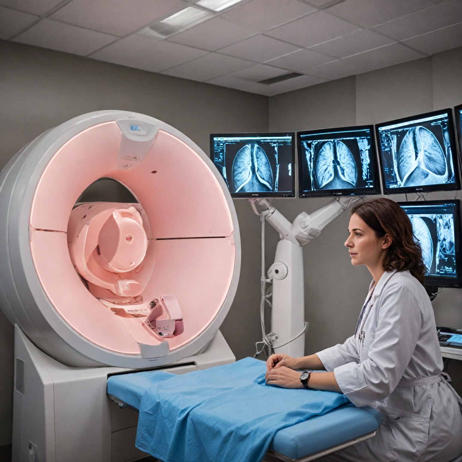
During the procedure, breast positioning and compression are conducted similarly to a standard digital mammogram. For each breast, two mammographic exposures are obtained in two different positions, resulting in a total of four images per breast and eight images for a complete CEM exam. Each exposure consists of a low-energy (LE) image resembling a conventional mammogram and a high-energy (HE) image, where the x-ray beam energy is tuned just above the k-edge of iodine to maximize attenuation by the contrast agent. The LE and HE images are processed by specialized software to generate an iodine-only image that highlights areas of contrast enhancement. Cancerous lesions typically exhibit greater enhancement, appearing as bright white regions against a darker background of normal tissue, aiding in lesion detection.
The acquisition of the LE and HE images is rapid, typically within a second each, with less than one second between acquisitions. The entire imaging sequence must be completed within approximately 10 minutes after contrast injection to avoid significant washout of the contrast agent from the breast tissue. Patient repositioning and gantry movements are performed at a moderate pace to ensure all views are obtained within this timeframe.
A radiologic technologist or mammographer is responsible for patient positioning and operation of the mammography equipment, while a radiologist analyzes the resulting images and communicates findings to the referring healthcare provider. The procedure is designed to minimize patient discomfort and anxiety, and the use of contrast enhancement improves visualization of breast lesions, particularly in women with dense breast tissue where traditional mammography may have reduced sensitivity.
Limitations, Risks, and Challenges
Despite advances in
Patient Advocacy and Legislative Impact
Patient advocacy and legislative efforts have played a crucial role in the rapid adoption of digital breast tomosynthesis (DBT) as a supplemental screening tool for women with dense breasts. One of the key driving forces behind the diffusion of DBT in U.S. community practices has been the breast density legislation movement, which has heightened awareness among patients and providers about the limitations of traditional mammography in detecting cancer in dense breast tissue. Such advocacy has increased patient demand for supplemental screening options, contributing to the widespread integration of adjunct tomosynthesis, which has shown potential to reduce false-positive rates and shift breast imaging practices towards higher volumes of screening rather than diagnostic workups.
Legislative mandates, including updates to the Mammography Quality Standards Act (MQSA) by the U.S. Food and Drug Administration, now require mammography facilities to inform patients whether they have dense breast tissue, due to its known association with increased breast cancer risk and the masking effect it has on mammograms. This transparency aims to empower patients with dense breasts—who constitute nearly half of women aged 40 and older undergoing mammography—to engage in informed discussions about supplemental screening options. Patient advocacy groups have been instrumental in promoting such legislation, advocating for policies that improve early detection and reduce the harms associated with false-positive results in this population.
The combined effect of patient-driven demand and legislative measures has accelerated the clinical uptake of DBT, with evidence supporting its improved screening outcomes for the majority of women, although benefits remain limited for a subset of women with extremely dense breasts. Ultimately, advocacy and legislation have enhanced awareness, informed patient choice, and influenced clinical practice patterns, helping to address the challenges posed by dense breast tissue in breast cancer screening.
Future Directions and Developments
The future of breast cancer screening for women with dense breasts is marked by ongoing research and technological advancements aimed at improving detection accuracy and reducing false positives. One promising area is the integration of digital breast tomosynthesis (DBT) with other imaging modalities and adjunct techniques. Trials such as MERIT and C-MERIT are currently exploring the combined use of blood tests, detailed patient history, and contrast-enhanced mammography (CEM) alongside conventional screening mammograms to detect breast cancer earlier, especially in women with dense breast tissue. Similarly, the CMIST trial is comparing CEM with DBT to evaluate the most effective screening method for this population.
Contrast-enhanced mammography employs iodine-based contrast agents, similar to those used in CT scans, which enhance the visualization of cancerous areas by increasing image contrast in dense breast tissue. This technique may provide superior imaging compared to traditional 2D or 3D mammography and is also useful in assessing the extent of cancer and monitoring response to chemotherapy. However, technical considerations such as the use of high-energy x-ray beams tailored to maximize iodine attenuation result in enhanced images that may not be diagnostic by themselves but improve overall assessment when combined with low-energy images.
Another significant development is the refinement of DBT technology. Current DBT systems acquire multiple low-dose 2D projections across an arc, reconstructing them into slice images that provide a 3D representation of the breast, improving lesion conspicuity and potentially increasing invasive cancer detection rates in women with dense breasts. Despite these benefits, recruitment for large-scale trials like the Tomosynthesis Mammographic Imaging Screening Trial has faced challenges as more hospitals adopt DBT, complicating study design and data collection.
Cost-effectiveness and clinical effectiveness remain critical factors under evaluation. A recent systematic review comparing DBT, digital mammography, and ultrasound for women with dense breasts and additional risk factors found that DBT might offer higher accuracy in symptomatic women or those recalled after screening. These findings support the growing advocacy for DBT adoption driven by breast density legislation and increased patient demand for supplemental screening modalities that reduce false-positive rates and shift practice toward higher screening volumes.
Looking forward, breast cancer screening guidelines continue to evolve. Organizations such as the American Cancer Society endorse the choice between 2D and 3D mammography based on individual risk and clinical judgment, emphasizing that financial barriers should not impede access to either modality. The combination of advanced imaging techniques like DBT and CEM, along with personalized risk assessment tools, holds promise for enhancing early detection and improving outcomes for women with dense breasts.
The content is provided by Jordan Fields, Lifelong Health Tips