Summary
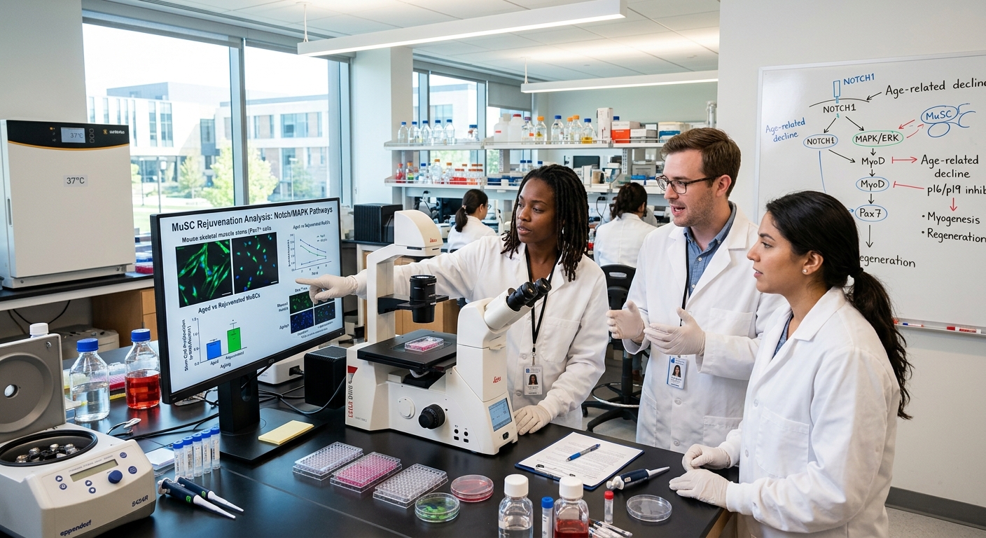
Unlocking Youth: How Older Muscle Stem Cells Can Regain Their Vitality — But There’s a Twist explores the biology, decline, and potential rejuvenation of muscle stem cells (MuSCs), also known as satellite cells, which are essential for skeletal muscle repair and regeneration throughout life. These cells reside in a specialized niche and remain mostly quiescent until activated by injury or stress. However, aging leads to a progressive decline in their number and regenerative function, contributing to sarcopenia—a condition marked by muscle loss, frailty, and reduced quality of life in the elderly. This decline results from a complex interplay of intrinsic cellular changes, such as DNA damage and altered signaling pathways, and extrinsic factors, including systemic inflammation and niche deterioration.
Research over recent decades has revealed that the impaired function of aged MuSCs is not irreversible. Experimental interventions, including exposure to a youthful systemic environment through heterochronic parabiosis and modulation of key signaling pathways like Notch and MAPK/ERK, have demonstrated significant restoration of regenerative capacity in aged muscle stem cells. Additionally, metabolic processes such as autophagy decline with age but can be rejuvenated, further enhancing MuSC function. These findings underscore the critical influence of both intrinsic cellular mechanisms and the extracellular niche in determining muscle stem cell vitality and open avenues for therapeutic strategies aimed at combating age-associated muscle degeneration.
Despite these promising advances, challenges remain. The rejuvenation of aged MuSCs is complicated by methodological issues in assessing stem cell function, potential trade-offs between stem cell survival and performance, and the existence of an irreversible senescence threshold in geriatric muscle stem cells. Furthermore, some molecular alterations observed in aged MuSCs may be protective adaptations rather than purely detrimental changes, adding complexity to therapeutic targeting. Effective treatments will likely require combined approaches that address both cell-intrinsic defects and the aged microenvironment, including niche remodeling and senescent cell clearance.
Looking forward, the integration of bioengineering, cell therapy, and molecular modulation holds great promise for restoring youthful function to aged muscle stem cells. Strategies under investigation include targeted manipulation of signaling pathways, epigenetic and metabolic reprogramming, and development of biomaterials that mimic youthful niche conditions to enhance stem cell engraftment and expansion. Such multidisciplinary efforts aim not only to treat sarcopenia and muscle injuries but also to improve overall muscle health and resilience in the aging population, marking an exciting frontier in regenerative medicine.
Background
Skeletal muscle possesses a remarkable regenerative capacity largely attributed to resident muscle stem cells, known as satellite cells or muscle stem cells (MuSCs), which serve as myogenic precursors essential for muscle repair and regeneration throughout life. These satellite cells reside in a specialized niche and remain mostly quiescent until activated by injury or stress, upon which they proliferate, differentiate, and fuse to existing fibers or form new ones to maintain muscle homeostasis.
However, aging is accompanied by a progressive decline in muscle mass and function, a condition termed sarcopenia, which contributes to reduced mobility, frailty, and diminished quality of life in elderly individuals. This decline is characterized not only by a loss in the quantity and regenerative capacity of MuSCs but also by alterations in their surrounding niche and systemic environment. Aging impairs the ability of satellite cells to maintain quiescence and self-renewal, resulting in aberrant activation and reduced regenerative potential.
The mechanisms underlying age-related muscle deterioration are multifactorial and include intrinsic impairments within the stem cells themselves as well as extrinsic changes in the aged local and systemic milieu. These extrinsic factors encompass chronic inflammation, fibrogenic signaling, mitochondrial dysfunction, and altered intercellular communication pathways such as Delta-Notch, Wnt, FGF, and others. Notably, studies in animal models have demonstrated that systemic factors from young organisms can rejuvenate aged muscle regeneration, highlighting the critical role of the microenvironment in modulating MuSC function.
Despite advances in understanding these processes, the molecular identities and regulatory mechanisms of human muscle stem and progenitor cells remain incompletely characterized, particularly in the context of aging and disease. Emerging transcriptomic analyses from young and elderly human muscle biopsies have begun to reveal distinct cellular populations and age-associated changes, providing deeper insights into the drivers of sarcopenia and frailty.
Molecular and Cellular Mechanisms Underlying Decline in Older Muscle Stem Cells
Muscle stem cells (MuSCs), also known as satellite cells, experience a multifaceted decline in function during aging, driven by both intrinsic cellular changes and alterations in their systemic and niche environments. This decline manifests in reduced regenerative capacity and impaired muscle repair, contributing to sarcopenia and frailty in aged organisms.
Intrinsic Cellular Alterations
Aging MuSCs exhibit increased DNA damage, including single- and double-stranded breaks, which, if unresolved, induce cellular senescence through pathways such as the p53/p21^CIP1^ and p16^INK4a^-Rb axes. The accumulation of DNA damage triggers apoptosis and senescence, diminishing the stem cell pool and regenerative potential. Alongside genomic instability, mitochondrial dysfunction and reduced autophagic activity are prominent features in aged MuSCs, leading to impaired energy metabolism necessary for activation and proliferation.
Furthermore, aged muscle stem cells show altered signaling dynamics. Notably, there is a decline in mitogen-activated protein kinase (MAPK)/phosphorylated extracellular signal-regulated kinase (pERK) signaling, which is crucial for activating Notch signaling pathways essential for MuSC function. Concurrently, increased activity of the p38αβ MAPK and JAK/STAT3 pathways in quiescent aged satellite cells has been observed, alongside derepression of the p16^INK4a^ locus at advanced ages, collectively contributing to cellular senescence and loss of regenerative capacity.
Aging also alters the balance between quiescence and activation in MuSCs. The FoxO3–Notch–Pax7–MyoD axis has emerged as a key regulatory pathway for maintaining or re-establishing quiescence during regeneration, with FoxO3 promoting Notch receptor expression and thus quiescence re-entry. Post-transcriptional regulation is also involved, where factors such as CPEB1 and Lin28 facilitate translation of mitochondrial oxidative phosphorylation (OXPHOS) transcripts, while microRNAs like miR-1/133α negatively regulate key myogenic transcripts, influencing the activation and proliferation capacity of MuSCs.
Another intrinsic factor is the age-associated increase of NDRG1 protein in muscle stem cells, acting as a molecular brake by suppressing mTOR signaling, which normally promotes cell growth and activation. Elevated NDRG1 levels lead to slower MuSC activation but enhance survival, suggesting a cellular “survivorship bias” that favors resilient but functionally compromised stem cells in aged muscle.
Extrinsic and Niche-Related Influences
The systemic environment and local niche also play crucial roles in MuSC aging. Age-associated changes in circulating factors include elevated levels of transforming growth factor-beta (TGFβ) family members, Wnt, and oxytocin, alongside decreased fibronectin availability in the stem cell niche. These changes impair β1-integrin and FGF2-induced ERK signaling, which are important for MuSC activation, proliferation, and self-renewal. Additionally, the aged niche shows altered interactions among diverse cell types involved in muscle regeneration, such as inflammatory cells and fibro-adipogenic progenitors, affecting the orchestrated regenerative process.
Studies indicate that extrinsic environmental factors and intrinsic cellular alterations act synergistically to drive MuSC dysfunction with age. While some satellite-cell decay can be reversed by exposure to a “youthful” systemic environment, other intrinsic mechanisms, such as epigenetic changes and senescence, persist despite environmental rejuvenation, highlighting the complexity of aging in muscle stem cells.
Impact of Autophagy and Protein Homeostasis
Autophagy, a critical process for cellular quality control and energy supply during MuSC activation, declines with age. Reduced autophagic flux leads to accumulation of damaged proteins and organelles, promoting cellular senescence and impairing regeneration. Restoration of autophagy in aged MuSCs has been shown to rescue regenerative potential, underscoring its importance in maintaining stem cell vitality during aging.
Markers Distinguishing Aged and Young Muscle Stem Cells
Muscle stem cells (MuSCs), also known as satellite cells, exhibit distinct molecular and functional profiles that vary with age, reflecting their altered regenerative capacity in aged tissues. A critical challenge in muscle biology is identifying reliable markers that differentiate young, highly regenerative MuSCs from their aged, functionally impaired counterparts.
One key observation is the shift in MuSC subpopulations during aging. Young MuSC pools are enriched for cells with low MYF5 expression (MYF5^Low), which are associated with long-term self-renewal capabilities. In contrast, aged MuSC pools are dominated by MYF5^High cells, indicating a reduction in the more stem-like subset with age. Additionally, single-cell RNA sequencing (scRNA-seq) analyses have revealed transcriptionally distinct Pax7-expressing MuSC subpopulations: Pax7^High cells, enriched in stemness-related genes and preferentially associated with glycolytic myofibers, and Pax7^Low cells, which express markers of myogenic differentiation. Aging alters the balance between these populations, impacting regenerative potential.
Surface marker expression also changes with age. MuSCs with higher levels of CD34 exhibit reduced differentiation propensity, linking CD34 expression levels to functional heterogeneity within the MuSC compartment. However, some canonical markers such as Pax7 and α7 integrin do not show significant differences in expression between young and old satellite cells in mice, suggesting that not all traditional markers reflect age-related functional changes.
Aging also influences senescence-associated markers in MuSCs. The proportion of satellite cells exhibiting elevated β-galactosidase (βgal) activity increases with age, serving as a senescence marker within myofibers and indicating a decline in satellite cell functionality. Importantly, this increase in βgal expression appears to be regulated post-transcriptionally, as endogenous Myf5 gene activity remains largely unchanged with age.
Intrinsic signaling alterations contribute further to the functional divergence between young and aged MuSCs. Elevated activity of pathways such as p38αβ mitogen-activated protein kinase (MAPK), JAK/STAT3, and TGF-β in aged quiescent satellite cells leads to impaired activation, proliferation, and self-renewal capacities. These signaling changes also influence the expression and activity of transcription factors like YY1, which regulates gene expression and metabolism in MuSCs, and whose deletion impairs regenerative responses.
Moreover, systemic and niche-derived extrinsic factors modulate MuSC aging phenotypes. Altered levels of fibronectin, Wnt, fibroblast growth factor-2 (FGF-2), and apelin in the aged muscle microenvironment diminish MuSC function and contribute to the observed phenotypic shifts. These extrinsic cues affect β1-integrin interactions and downstream ERK signaling, thereby modulating MuSC behavior during regeneration.
Experimental and Therapeutic Strategies to Rejuvenate Aged Muscle Stem Cells
Aged muscle stem cells (MuSCs), also known as satellite cells, exhibit diminished regenerative capacity due to both intrinsic cellular changes and alterations in their systemic and niche environments. To counteract this decline, multiple experimental and therapeutic strategies have been developed, targeting the complex interplay of molecular signaling pathways, metabolic processes, and niche interactions that govern MuSC function.
One promising avenue involves modulation of signaling pathways critical to MuSC activation and self-renewal. Notch signaling plays a pivotal role in promoting muscle regeneration by activating satellite cells and driving their proliferation. Activation of Notch signaling has been shown to restore regenerative potential in aged muscle, whereas its inhibition impairs regeneration in young muscle tissue. Furthermore, mitogen-activated protein kinase (MAPK)/extracellular signal-regulated kinase (ERK) signaling, which declines with age, is important for Notch activation in human muscle stem cells, suggesting that restoring MAPK/pERK activity could rejuvenate aged MuSCs. Other pathways such as p38 MAP kinase, β-catenin, Stat3, FGF, and SMAD are dysregulated in aged MuSCs and represent potential therapeutic targets.
Metabolic regulation is another key factor in stem cell rejuvenation. Autophagy, a cellular recycling process, is crucial for the activation and proliferation of MuSCs, serving as a temporary energy source. Its decline with age leads to MuSC senescence and impaired muscle regeneration. Strategies aimed at restoring autophagy, such as exercise-induced activation of SIRT1, have shown potential in ameliorating dysfunctional autophagy in satellite cells, although exercise alone does not fully prevent age-related muscle defects. Additionally, transcriptional and post-transcriptional regulators like YY1 and microRNAs modulate gene expression and metabolism in MuSCs, influencing their regenerative capabilities.
Given the critical influence of the stem cell niche and systemic environment on MuSC function, therapeutic interventions also focus on modifying these extrinsic factors. Heterochronic parabiosis experiments—where the circulatory systems of young and aged mice are surgically joined—have demonstrated that exposure to young systemic factors can rejuvenate aged satellite cells and enhance their regenerative response following injury. Conversely, aged tissues can rapidly induce aging phenotypes in young MuSCs, highlighting the necessity of rejuvenating both intrinsic cell properties and the local milieu to achieve effective regeneration. Approaches that combine cell-intrinsic rejuvenation with modulation of the niche, such as extracellular matrix remodeling, elimination of senescent cells, or administration of paracrine factors, have been proposed as comprehensive strategies to restore muscle stem cell function.
Cell therapy using autologous transplantation of potent muscle stem cells offers a tantalizing possibility for treating congenital muscle diseases like Duchenne Muscular Dystrophy and muscle injuries. However, the aged niche’s dysfunction poses challenges to the efficacy of such therapies, underscoring the importance of a dual approach addressing both stem cells and their environment.
Emerging techniques including epigenetic modulation, genetic enhancement, in vivo partial reprogramming, and metabolic manipulation are under investigation to directly rejuvenate MuSCs. Targeted manipulation of signaling networks governing quiescence, activation, and self-renewal may unlock unique opportunities for muscle regenerative medicine, paving the way for interventions that restore youthful function to aged muscle stem cells.
Experimental Evidence
Recent experimental studies have provided compelling evidence that aged muscle stem cells, also known as satellite cells, can regain their vitality and restore muscle function under certain conditions. One key approach has involved transplanting treated old muscle stem cells back into elderly mice, which led to the restoration of muscle strength to levels comparable to those of younger animals. This rejuvenation was observed across multiple cellular hallmarks of aging, highlighting the broad potential for reversing age-associated decline in muscle regenerative capacity.
A critical factor influencing these outcomes is the methodology used to assess satellite cell function. It has been proposed that isolating satellite cells followed by transplantation may exacerbate intrinsic aging-related changes, thus altering their natural function and contributing to discrepancies among different studies. This suggests that the environment and procedure employed significantly impact the observed regenerative potential of aged satellite cells.
Systemic factors have also been implicated in muscle stem cell rejuvenation. The classical parabiosis technique, which surgically joins the circulatory systems of young and old mice, demonstrated that exposure to young blood can restore satellite cell activation and enhance muscle regeneration in aged animals. These findings support the existence of circulating “rejuvenation” factors capable of modulating muscle repair mechanisms in older organisms.
Further supporting this notion, regular exercise has been shown to restore youthful regenerative capacity in aged muscle stem cells, indicating that lifestyle interventions may also play a role in maintaining stem cell function with age. Molecular studies from UCLA suggest that certain age-related molecular changes in muscle stem cells may be protective rather than solely detrimental, adding complexity to our understanding of stem cell aging.
In addition to murine models, efforts are underway to translate these findings to human muscle stem cells. Strategies involving the isolation, rejuvenation, and expansion of human muscle stem cells using bioengineering and small molecule treatments have shown promise. Transplantation of these rejuvenated cells into aged or atrophied
Challenges and Considerations
Rejuvenating aged muscle stem cells (MuSCs) to restore their regenerative capacity presents several challenges and important considerations. One significant issue arises from the methods used to assess the potential for young environments to rejuvenate aged satellite cells. Isolation and transplantation procedures may inadvertently exacerbate intrinsic alterations within aged MuSCs, thus influencing experimental outcomes and leading to discrepancies among studies. This suggests that the context and methodology of stem cell evaluation critically affect interpretations of their regenerative potential.
Furthermore, enhancing aged stem cell function is not without risks. As emphasized by Rando, improvements in one aspect of stem cell activity may entail unintended consequences or trade-offs, indicating a delicate balance between stem cell survival and performance during aging. This complexity underscores the need for careful examination of molecular mechanisms governing stem cell behavior to avoid adverse effects.
Another key consideration is the timing of therapeutic interventions. Evidence indicates that the muscle stem cell pool enters a state of irreversible senescence at a geriatric age threshold, marking a point of no return beyond which rejuvenation may be ineffective. Therefore, treatments aiming to restore endogenous stem cell function should ideally be applied before this critical juncture to maximize efficacy.
At the molecular level, DNA damage accumulation in MuSCs during aging contributes to functional decline. Persistent single- or double-stranded DNA breaks can trigger cellular senescence through pathways such as p53/p21^CIP1^, leading to apoptosis and reduced regenerative potential. Pharmacologic activation of p53 has been shown to suppress DNA damage and ameliorate cell death in aged MuSCs, suggesting a potential therapeutic target. However, the interplay between DNA damage responses and stem cell function is complex, and some molecular changes observed in aged cells may be protective rather than solely detrimental.
Lastly, the multifaceted nature of stem cell aging involves intrinsic factors, such as epigenetic and metabolic alterations, as well as extrinsic influences from the stem cell niche. Strategies for rejuvenation must therefore consider both cell-intrinsic and niche-related components, including modulation of signaling pathways, extracellular matrix remodeling, elimination of senescent cells, and delivery of bioengineered molecules to restore SC–niche interactions. Balancing these approaches while mitigating potential risks remains a major challenge in developing effective therapies to counteract muscle stem cell exhaustion and age-related regenerative decline.
Future Directions
Recent advances in understanding the decline of muscle stem cell (MuSC) function with age have opened several promising avenues for therapeutic intervention aimed at restoring muscle regenerative capacity. A key insight is the recognition that the MuSC pool undergoes an irreversible senescence state at geriatric age, indicating that rejuvenation therapies should ideally be applied before this critical threshold to preserve regenerative potential. Early intervention strategies may focus on maintaining the quiescent, highly regenerative MuSC subpopulations throughout life and expanding these relevant progenitor pools within the stem cell niche to reverse age-associated muscle deterioration.
One promising approach involves restoring the interactions between MuSCs and their niche through the delivery of bioengineered molecules designed to recapitulate the native biochemical and biophysical environment of young muscle tissue. These biomaterials could address the challenges posed by direct cell delivery methods and enhance therapeutic outcomes in muscle diseases and injuries, including volumetric muscle loss (VML). By mimicking the complex inflammatory microenvironment that supports in vivo MuSC expansion, it may be possible to sustain stem cell potency during in vitro culture and improve engraftment success.
Cell-based therapies continue to show great potential, as human muscle stem cells demonstrate precise homing to their appropriate niches and contribute functionally to muscle fibers without tumorigenic risks. However, intrinsic defects in aged MuSCs are not fully rescued by transplantation into a young environment, underscoring the need to identify and therapeutically target molecular alterations within aged cells themselves. Investigating the signaling pathways that regulate stem cell maintenance and function, as well as epigenetic and metabolic modulators, offers a rich landscape for developing rejuvenation strategies, including small molecules, genetic enhancements, and partial in vivo reprogramming.
Additionally, approaches targeting senescence, such as senolytic therapies aimed at selectively eliminating senescent cells, represent a complementary strategy to rejuvenate the muscle stem cell niche and improve tissue function. Integrating stem cell biology with immune modulation and metabolic reprogramming may further enhance muscle regeneration, as indicated by studies focusing on “stem cell-immune-muscle regeneration” regulatory modules.
The content is provided by Avery Redwood, Lifelong Health Tips