Summary
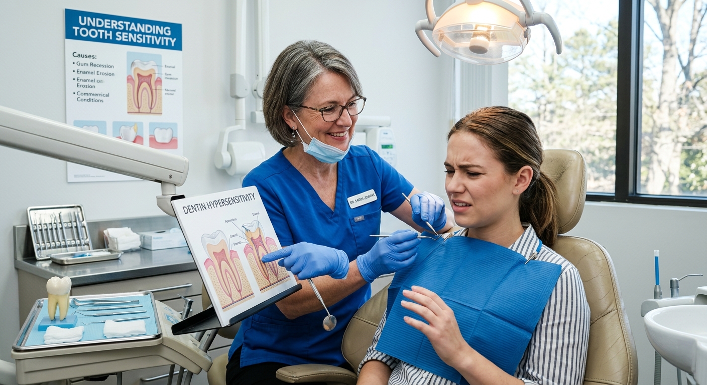
The Secrets Behind Sensitive Teeth: Understanding and Easing Your Discomfort explores the causes, biological mechanisms, symptoms, diagnosis, treatment, and prevention of tooth sensitivity, clinically known as dentin hypersensitivity (DH). This common dental condition affects a significant portion of the population, with prevalence estimates ranging from 8% to 57% worldwide, and is characterized by sharp, transient pain in response to thermal, chemical, or tactile stimuli. The discomfort arises primarily from the exposure of dentin—the tooth layer containing nerve endings—due to factors such as gum recession, enamel erosion, tooth decay, or dental procedures. Sensitive teeth can severely impact quality of life by restricting dietary choices and causing psychological distress.
At the core of dentin hypersensitivity is the hydrodynamic theory, which explains pain as a result of fluid movement within microscopic dentinal tubules that stimulates nerve endings in the dental pulp. Recent advances have elucidated complex cellular and molecular pathways involving odontoblasts, sensory neurons, and ion channels such as transient receptor potential (TRP) channels, contributing to pain perception and amplification. These insights have paved the way for novel treatments targeting the underlying mechanisms of sensitivity.
Management of sensitive teeth involves a multifaceted approach, including professional in-office treatments like fluoride varnishes and tubule-sealing agents, as well as at-home care featuring desensitizing toothpastes and lifestyle modifications to protect enamel and gums. Emerging therapies utilizing nano-hydroxyapatite and arginine-calcium carbonate formulations show promising results in reducing sensitivity. Preventive strategies focus on maintaining gentle oral hygiene, avoiding acidic and abrasive exposures, and regular dental check-ups to mitigate risk factors.
Despite advances, challenges remain in accurately diagnosing dentin hypersensitivity due to overlapping symptoms with other dental conditions and the reliance on patient self-reporting. Moreover, variability in treatment responses underscores the need for personalized care and further research. Addressing tooth sensitivity effectively not only alleviates pain but also prevents progression to more severe dental issues, underscoring its significance in oral health management.
Causes
Tooth sensitivity, also known as dentin hypersensitivity, can arise from a variety of underlying factors that expose or irritate the dentin layer of the tooth, which contains nerve endings. One of the primary causes is gum recession, often resulting from aggressive brushing, periodontal disease, trauma, or plaque and tartar buildup. When gums recede, the underlying dentin becomes exposed, making teeth more susceptible to sensitivity from stimuli such as hot, cold, or sweet substances.
Enamel erosion is another significant cause of tooth sensitivity. The enamel is the hardest substance in the human body, protecting the underlying dentin. However, repeated exposure to acids from foods and beverages, or from gastric reflux, can wear down this protective layer. As enamel thins, microscopic pores form, allowing stimuli to penetrate deeper into the tooth structure, which increases sensitivity and vulnerability to further damage. In later stages, enamel erosion can cause severe sensitivity and visible changes like discoloration and cupping on the tooth surface.
Cracked or fractured teeth can also lead to sensitivity. Damage to the tooth structure may expose the dentin or allow bacteria to infiltrate, causing irritation and pain. Similarly, tooth decay resulting from bacterial activity on enamel can break down the protective surface and expose sensitive areas beneath.
Other contributing factors include gum disease, which causes the gums to become swollen, infected, or to pull away from the teeth, exposing the roots and increasing sensitivity. Additionally, habits such as tooth grinding or clenching can wear down enamel, further exposing the dentin and exacerbating sensitivity.
Certain dental procedures, including fillings, cleanings, or whitening treatments, can cause temporary tooth sensitivity. Mouthwash products containing acidic ingredients may also worsen sensitivity, and individuals are often advised to seek gentler formulations.
Finally, temporomandibular disorders (TMD), involving the jaw muscles, joints, and associated nerves, may be linked to chronic facial pain and contribute to the sensation of tooth sensitivity in some cases. Overall, the multifactorial nature of sensitive teeth highlights the importance of identifying and addressing specific causes to manage and alleviate discomfort effectively.
Pathophysiology
Dentin hypersensitivity (DH) arises primarily from the exposure of dentin, which is normally covered by enamel on the crown and a thin layer of cementum on the root surface of the tooth. When these protective layers are lost or compromised, the underlying dentin becomes exposed to external stimuli, resulting in pain and discomfort. The dentin contains numerous microscopic tubules that extend from the outer surface to the dental pulp, where nerve endings are located. The sensitivity is largely attributed to the movement of fluid within these dentinal tubules in response to various mechanical, thermal, or osmotic stimuli.
The most widely accepted explanation for the mechanism behind DH is the hydrodynamic theory. This theory proposes that external stimuli cause a rapid movement of fluid through the dentinal tubules, which leads to distortion of the dental pulp tissue and activation of nerve endings within the pulp, particularly in the plexus of Raschkow. This neural stimulation results in the sensation of pain. The extent of sensitivity is closely related to the patency, number, and diameter of dentinal tubules; wider tubules significantly increase fluid flow due to the fourth-power relationship between tubule radius and fluid movement, leading to heightened pain responses.
At the cellular level, odontoblasts and myelinated sensory neurons extend processes into the dentinal tubules, penetrating roughly one-third of their length. This anatomical arrangement has led to the hypothesis that odontoblasts may function as primary sensory cells, communicating with adjacent free nerve endings to trigger pain signals. Various ion channels and receptors expressed by odontoblasts and intradental nerve fibers—including transient receptor potential (TRP) channels such as TRPV1—play critical roles in sensing thermal and chemical stimuli. Activation of these channels increases intracellular calcium concentration in odontoblasts, leading to the release of neurotransmitters like ATP and glutamate, which then stimulate adjacent nerve fibers.
Pain signaling is further modulated by the expression of neuropeptides such as substance P (SP) and calcitonin gene-related peptide (CGRP), which are involved in neurogenic inflammation and pain amplification within the pulp. The dental pulp is densely innervated by sensory fibers from the trigeminal ganglia, predominantly unmyelinated fibers, making it highly responsive to nociceptive stimuli. Inflammatory conditions like pulpitis increase the levels of endogenous agonists for TRP channels and sensitize these channels, exacerbating pain.
Additionally, purinergic signaling through P2X receptors, especially P2X3 expressed on trigeminal nerve fibers, is implicated in the transmission of pain associated with dentin hypersensitivity. ATP released from activated odontoblasts binds to these receptors, promoting nociceptive signaling. This interaction is often co-expressed with other nociceptive receptors, suggesting a complex network of pain modulation in dental tissues.
Symptoms
Tooth sensitivity typically manifests as sudden, sharp discomfort or pain in response to hot, cold, sweet, or acidic stimuli. Commonly, individuals experience aching or sharp sensations when consuming hot beverages like coffee, cold foods such as ice cream, or even during routine activities like brushing teeth. This discomfort arises when the underlying dentin layer becomes exposed, allowing external stimuli to trigger nerve responses within the tooth.
The pain associated with sensitive teeth can vary in quality and intensity. A-delta nerve fibers are primarily responsible for the sharp, quick pain experienced upon mechanical or thermal stimulation, whereas C-fibers respond to more intense or prolonged stimuli, contributing to dull or aching sensations often linked to pulpal inflammation. Approximately 75% of patients with dentin hypersensitivity report heightened pain responses to cold stimuli, which cause fluid movement away from the dentin-pulp complex, exacerbating the sensation.
Additional symptoms that may accompany sensitivity include gum recession and inflammation, which expose the tooth roots and increase vulnerability to external triggers. Patients might also notice changes such as swelling or a “pulling away” of the gums, indicating underlying gum disease or damage that contributes to sensitivity. Sensitivity can persist transiently after dental treatments like fillings, crowns, or bleaching, usually subsiding within 2–3 days; however, prolonged or worsening sensitivity beyond this period should prompt professional evaluation.
Diagnosis
Diagnosing dentin hypersensitivity (DH) can be challenging, as it is primarily a diagnosis of exclusion reached after ruling out other possible causes of dental pain. A thorough patient history and clinical examination are essential components of the diagnostic process. The clinical examination often involves pain provocation tests, such as blasting air from a dental instrument onto the sensitive area or gently scratching the dentin surface with a dental probe to elicit a pain response.
Practitioners frequently rely on patients’ spontaneous complaints of sensitivity to identify DH, reflecting a lack of consensus on a standardized clinical diagnostic method. This reliance on patient self-reporting suggests that the true prevalence of DH may be underestimated, as objective diagnostic tests are used less frequently. Additionally, differences in populations studied and diagnostic methodologies contribute to the wide reported incidence range of 4% to 74%.
Sensitive teeth are characterized by a significantly higher number and larger diameter of patent dentinal tubules compared to non-sensitive teeth, which contributes to the pain response when stimuli reach these tubules. The increased tubule diameter is particularly relevant because fluid flow within dentinal tubules is proportional to the fourth power of the radius, meaning that even a small increase in diameter can dramatically enhance fluid movement and sensitivity. This physiological basis supports the clinical findings observed during diagnosis.
Differential diagnosis is critical since conditions such as gum disease, cavities, cracked teeth, or non-carious cervical lesions (NCCLs) can mimic or contribute to symptoms of DH. Therefore, clinicians must carefully evaluate for other dental issues that may cause or exacerbate sensitivity before confirming a diagnosis of DH.
Treatment
Managing tooth sensitivity involves a combination of professional dental procedures and at-home care tailored to the underlying cause and patient preferences. Treatment options range from simple lifestyle modifications to advanced clinical interventions designed to reduce discomfort and protect exposed dentin or nerves.
Professional In-Office Treatments
Dentists may apply several in-office therapies to alleviate dentinal hypersensitivity (DH). Common approaches include fluoride varnishes, which strengthen enamel and provide a protective coating, as well as treatments with glutaraldehyde combined with hydroxyethyl methacrylate (HEMA) to seal dentinal tubules. Other clinical options include the use of dentin-bonding agents, arginine-calcium carbonate pastes, calcium phosphate therapies, and laser treatments, all aimed at occluding exposed dentinal tubules and reducing nerve irritation.
For cases where tooth sensitivity arises from dental damage or disease, restorative procedures such as dental bonding, sealants, fillings, or crowns may be necessary to cover exposed dentin and prevent further irritation. In severe instances, where nerve involvement causes persistent pain, root canal therapy can be employed to remove damaged nerve tissue and provide lasting relief.
At-Home Care and Lifestyle Adjustments
Effective management of tooth sensitivity often involves changes to oral hygiene routines. Using a soft-bristled toothbrush and brushing gently with fluoride-containing toothpaste helps minimize enamel erosion and gum recession, which are common contributors to sensitivity. Selecting desensitizing toothpastes with active ingredients such as potassium nitrate or stannous fluoride has been shown to block pain signals from the tooth surface to the nerve, providing gradual relief over time.
Patients are also advised to avoid behaviors that exacerbate sensitivity, including chewing ice, consuming acidic foods and beverages, and overusing abrasive teeth-whitening products. Additionally, rinsing with warm salt water can reduce inflammation and soothe gum tissue, offering temporary comfort.
Desensitizing Agents and New Developments
Recent evidence supports the effectiveness of desensitizing agents containing nano-hydroxyapatite (n-HAP) in both at-home and clinical settings for reducing DH. While these agents show promise, further long-term clinical trials are needed to establish definitive recommendations.
Other emerging options include toothpastes containing arginine and calcium carbonate combined with fluoride, which have demonstrated efficacy in providing rapid relief of dentin hypersensitivity compared to traditional potassium-based formulations. Over-the-counter products such as Crest Pro-Health Sensi-Stop Strips and topical pain relief creams like Orajel may also offer immediate, though temporary, relief for sensitive teeth.
Comprehensive Management and Preventive Care
Optimal treatment outcomes are typically achieved through a combination of professional care and consistent home maintenance. Patients are encouraged to maintain good oral hygiene practices, use fluoride mouth rinses as recommended, and schedule regular dental check-ups to monitor and manage sensitivity effectively. Addressing contributory factors such as gum disease, tooth grinding, and cavities promptly can prevent worsening sensitivity and improve overall oral health.
Because treatment responses may vary based on individual health conditions and sensitivities, consultation with a dental professional is essential to tailor the approach and ensure safe, effective management of tooth sensitivity.
Prevention
Preventing tooth sensitivity involves adopting proper oral hygiene practices and making specific lifestyle adjustments aimed at protecting tooth enamel and gum health. Maintaining good oral hygiene is fundamental; brushing teeth twice daily with fluoride toothpaste and flossing daily help remove plaque and prevent gum disease, which can contribute to sensitivity. Using a soft-bristled toothbrush is recommended to minimize enamel abrasion and gum recession, both of which expose dentin and increase sensitivity.
Protecting tooth enamel is key to preventing sensitivity. Enamel serves as the hard outer layer shielding teeth, so avoiding its erosion is essential. This can be achieved by steering clear of acidic foods and drinks, which can wear down enamel over time. After consuming acidic substances, drinking water or using a straw can help reduce acid contact with teeth and balance oral pH levels. Additionally, quitting smoking and addressing habits like teeth grinding or clenching, often with the aid of a mouthguard, can prevent enamel wear and reduce sensitivity.
Gentle flossing is advised to prevent gum recession, and it is important not to brush too hard or too often, as aggressive brushing techniques can exacerbate enamel loss and gum damage. Regular dental check-ups enable early detection and management of sensitivity causes, allowing dentists to recommend tailored preventive measures or treatments if needed.
Lifestyle modifications also include avoiding chewing ice and limiting or cautiously using teeth-whitening products, which may aggravate sensitivity. While numerous over-the-counter desensitizing products exist, their clinical efficacy varies, and professional guidance is recommended for persistent sensitivity. Overall, a combination of diligent oral care, mindful habits, and professional consultation forms the cornerstone of effective prevention of sensitive teeth.
Impact on Quality of Life
Tooth sensitivity, often characterized by sudden and sharp pain when consuming hot, cold, or sweet foods and drinks, can significantly affect an individual’s daily life and well-being. The condition, known as dentin hypersensitivity, has a high prevalence worldwide, affecting between 8% and 57% of the general population, and can cause considerable psychological distress. This discomfort not only interrupts the simple enjoyment of eating and drinking but can also lead to a reduced overall quality of life due to persistent pain and avoidance behaviors.
Individuals with sensitive teeth may experience frequent discomfort that influences their dietary choices and social interactions, potentially leading to emotional and mental health challenges. The ongoing pain can cause anxiety around meals and diminish the pleasure derived from food and beverages, which are central aspects of daily living. Moreover, the condition’s unpredictability and intensity contribute to stress and frustration, emphasizing the need for effective management strategies.
Addressing tooth sensitivity is crucial not only to alleviate pain but also to prevent the escalation of underlying dental problems that may exacerbate discomfort and further impair quality of life. While reducing sensitivity provides relief, it is important to identify and treat the root causes
Research and Advances
Dentin hypersensitivity (DH) has been extensively studied to better understand its underlying mechanisms and improve clinical management. The predominant explanation for the pain experienced in DH is the hydrodynamic theory, which attributes sensitivity to the movement of fluid within the dentinal tubules. This fluid movement, often triggered by thermal, mechanical, or osmotic stimuli, activates baroreceptors leading to neural discharge and pain sensation. The mechanism is comparable to the activation of neural fibers around hair follicles when stimulated.
Recent research has expanded the understanding of molecular signaling involved in DH. Odontoblasts, cells lining the dental pulp, have been shown to communicate with trigeminal ganglion (TG) neurons through the release of signaling molecules such as ATP and glutamate. Mechanical stimulation of odontoblasts increases intracellular calcium (Ca2+) levels, triggering the release of these molecules which act on purinergic and metabotropic glutamate receptors on adjacent nerve fibers, contributing to pain signaling. Furthermore, bacterial components can directly activate nociceptive neurons, enhancing hypersensitivity through pathways involving toll-like receptor 4 (TLR4) and transient receptor potential (TRP) channels.
In terms of treatment advances, desensitizing agents containing nano-hydroxyapatite (n-HAP) have shown efficacy in reducing dentin hypersensitivity in both at-home and in-office settings. Meta-analyses suggest these agents perform better than placebo or other desensitizers, although further long-term studies are needed to confirm their effectiveness definitively. Additionally, diamine silver fluoride has been recognized as a clinically effective and safe tooth desensitizer with regulatory clearance, providing a viable option for managing DH.
Besides clinical treatments, numerous desensitizing toothpastes are available commercially. Patients are often encouraged to try different brands to identify which product best alleviates their sensitivity symptoms. Overall, ongoing research continues to refine the biological understanding and therapeutic approaches for dentin hypersensitivity, aiming to improve patient outcomes.
The content is provided by Jordan Fields, Lifelong Health Tips